Advanced Cavity Diagnosis
In our office we have some extra technology for diagnosing and monitoring early cavities.
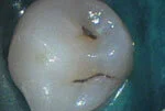
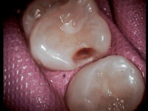
Pit and groove cavities
Conventional dentistry involved poking in the grooves of the teeth with a small sharp instrument called an explorer. If the groove was mushy or sticky, it was called a cavity.
By the time you can find a cavity in this fashion, it’s pretty advanced, and usually needs local anesthetic and significant decay removal.

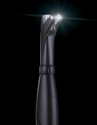
In our office we have a device called a Diagnodent, a laser sensor that can measure decay in grooves with much greater sensitivity.
We can tell the difference between simple stain and early decay. We can record and monitor early lesions that are stable, instead of trying to guess whether they need treatment.
When lesions progress, we can find the perfect “sweet spot” when they have just progressed enough that they need early treatment, but can be treated with microdentistry techniques that do not require anesthetic or “drilling”. This instrument helps us avoid sealing or treating teeth when unnecessary, as well as letting us know early when treatment is necessary.

Groove repaired with microfilling- no anesthetic or “drilling” needed, using Bioactive materials.
Cavities between teeth
There is no x-ray exposure, just an infrared camera.
Conventional dentistry only had one method of finding and tracking decay between teeth- accurately taken x-rays. Over the years the x-ray exposures have gone down and quality has gone up, and it with our second-generation digital sensors and narrow collimator shields we have the lowest exposures currently possible, but now there is a new alternative.
We got a Carivue system in 2015. This infrared camera allows us to turn the tooth transparent, so we can see right through the enamel to check for decay between teeth.

Decay appears dark while the enamel is transparent.

This tooth looks fairly normal.
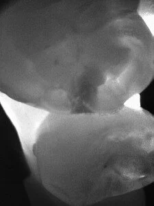
Decay visible on Carivue.

Decay is definitely present.

Final filling.
Like the Diagnodent, this lets us monitor early lesions, and lets us know when or if they get just big enough to need treatment. There is no health risk in doing this exam frequently.
It’s excellent for monitoring early cavities between teeth, so we can tell when to heal them medically or treat them with microdentistry. It’s also great for pregnant patients, kids with small mouths who can’t handle a digital x-ray, patients with a gag reflex, or patients who can’t have x-rays for medical reasons.
We have found it to be more accurate than x-rays for finding decay in unfilled teeth. Unfortunately, it can’t see through plastic fillings, gold, porcelain, titanium or bone.
Note: Insurance companies gladly cover x-rays, but even though an examination with diagnostic imaging is safer and less expensive, they don’t all cover the small fee yet.